Magnetic Resonance Imaging (MRI)

The MRI arrived at STEGH in July, 2024! Read more about it's arrival.
Click here to access the physician referral form.
What is an MRI?
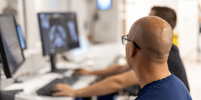
Magnetic Resonance Imaging (MRI) is a non-invasive medical imaging exam that helps radiologists to diagnose a variety of diseases including cancer. MRI uses a powerful magnetic field, radio frequency pulses, and a computer to produce detailed pictures of organs, soft tissues, bone, and other structures inside the body. In some cases, contrast material may be used during the MRI scan to show certain structures more clearly. Unlike CT scanning or general x-ray studies, no ionizing radiation is involved with an MRI.
In many cases, MRI gives different information about structure in the body than can be seen with an X-ray, ultrasound, or computed tomography (CT) scan. MRI also may show problems that cannot be seen with other imaging methods. This information is then sent to a computer which processes all the signals and generates it into an image. The final product is a 3-D image representation of the area being examined. These images can then be displayed and examined on a computer monitor, transmitted electronically, printed, or copied to a CD.
FAQ
Click here for more information about:
- Preparing for an MRI
- What should I wear?
- I'm claustrophobic, will I be able to have a scan?
- Do I need to have my creatinine level performed before my exam?
- What if I might be pregnant?
- What happens during an MRI?
- After your MRI
- How will I know the results of my MRI?
Safety first!
Click here for a list of items we need to know about in order to keep you safe.
Reference: Ontario Association of Radiologists


